
01 Lateral Pterygoid
Step 1
Understanding the Problem
This module teaches the release of the Lateral Pterygoid, a deep muscle of the jaw that controls forward movement of the mandible and stabilizes the temporomandibular joint. When this muscle enters spasm, it can contribute to jaw pain, clicking of the TMJ, headaches, and facial tension.
Step 2
The Clinical Problem


The mandibular nerve passing through the belly of the lateral pterygoid, close to where it exits the skull.


A client arrives complaining of jaw pain, clicking in the temporomandibular joint, headaches near the temples, or difficulty opening the mouth fully. Despite treatment of the masseter and temporalis muscles, the symptoms persist.
In many cases the underlying cause lies deeper within the jaw, where the lateral pterygoid muscle has entered spasm and is directly pinching the mandibular nerve which passes through its belly.
When in spasm it pinches this nerve and causes the extreme pain reported with TMJ.
It also tilts the body of the sphenoid bone forward.
The ends of this bone are locked in with the cranial bones that surround it in the temple. In its body it is flexible like a knife blade. The sella tursica can actually tilt forward and backward a bit. This small amount of movement is enough to depress the anterior edge of the sphenoid bone making it almost palpably flush with the palatine bone. This movement, if we focus on the back of the sella tursica pushes the sella tursica into the pituitary gland, by design. It is how the body squeezes pituitary hormones out of the pituitary gland. This explains why a yawn feels the way it does. The lateral pterygoid contracts, tilts the "backseat" of the sella tursica to tilt forward into the pituitary gland and then release. When the lateral pterygoid is in spasm, this becomes stuck and the pituitary gland becomes compressed all of the time. This causes disruptive dreams or nightmares. This causes many rippling side-effects related to hormones, since the pituitary gland is the master hormone gland of the body, connected neurally directly to the brain.
This release though technically challenging is the key to profound health benefits. Release of the mandibular nerve and freedom from TMJ pain is only the beginning.
Step 3
Why This Happens
The Root Cause of Muscle Spasm (Lateral Pterygoid)

Most chronic pain and dysfunction in the jaw and head originates from muscle spasm rather than structural damage.
In this modality, the primary muscle involved is the lateral pterygoid, which is frequently strained by:
• clenching or grinding the teeth (bruxism)
• stress-related jaw tension
• excessive chewing or gum use
• poor jaw alignment or posture
When the lateral pterygoid becomes fatigued or overused, it can enter a state of protective contraction, where the muscle fibers remain shortened and resist lengthening.
Over time, this spasm creates several problems:
• accumulation of static electrons
• reduced circulation through the tissue
• accumulation of metabolic waste
• inability to produce sufficient ATP
• impaired distribution of electrolytes
• compression within the temporomandibular joint (TMJ)
• restricted jaw movement
• referred pain into the face, temple, and head
The lateral pterygoid plays a critical role in controlling the position of the jaw and the TMJ disc. When it is in spasm, it can pull the disc forward, contributing to clicking, locking, and dysfunction of the jaw.
The body compensates by recruiting surrounding muscles of mastication, spreading tension throughout the face and head.
To resolve this, we identify the lateral pterygoid as the primary muscle in spasm, determine the activity that caused the fatigue, and remove that stress.
Patients must avoid aggravating behaviors such as clenching or excessive chewing for approximately six weeks after release.
Releasology restores the muscle’s ability to relax, allowing normal jaw mechanics and reducing facial tension and pain.
Step 4
Anatomy
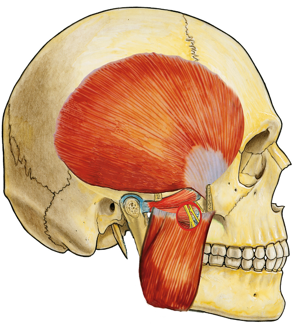
This modality focuses on the lateral pterygoid, the most influential muscles in the body for jaw mechanics, jaw pain, pituitary function, eyesite, vibrato for singers, nightmares or unsettling dreams and overall cranial dynamics. It is the keystone of the skull in many ways.
Key anatomical features include:
• Origin: sphenoid bone (greater wing and lateral pterygoid plate)
• Insertion: neck of the mandible and TMJ disc
• Fiber direction: horizontal
Function:
• protracts the mandible
• assists in opening the jaw
• stabilizes and positions the TMJ disc
The Mandibular Nerve Relationship
The mandibular branch of the trigeminal nerve (CN V3) exits the skull and passes directly through the region of the lateral pterygoid.
When the lateral pterygoid enters spasm:
• it compresses the mandibular nerve within its fibers
• this can mechanically restrict signaling through the trigeminal system
• all muscles of mastication may reflexively contract
• sensory branches from the teeth, jaw, and face may send pain signals
This explains why what is commonly diagnosed as TMJ pain is often not a joint issue at all, but a deep muscular compression of the mandibular nerve.
In many cases, the lateral pterygoid is the primary and sole cause of these symptoms.
The Role of the Sphenoid Bone
The lateral pterygoid originates from the sphenoid bone, a central bone of the skull that supports:
• the cranial base
• the eye structures
• the pituitary gland (within the sella turcica)
Although traditionally described as fixed, clinical palpation reveals that the sphenoid can exhibit subtle positional shifts.
When the lateral pterygoid is in spasm:
• it draws the sphenoid forward through its origin point
• the body of the sphenoid tilts slightly anteriorly
• this movement can be palpated internally at the junction with the palatine bone along the midline
Even a small shift (approximately 1 mm) can have noticeable effects.
Sella Turcica and Pituitary Relationship
At the center of the sphenoid lies the sella turcica, which houses the pituitary gland.
As the sphenoid tilts forward:
• the posterior aspect (“backsheet”) of the sella turcica may press against the pituitary
• this mechanical interaction may influence pituitary activity
This relationship is observed in Releasology as a possible explanation for:
• hormonal fluctuations
• vivid dreams or nightmares
• systemic changes following release
Yawning may represent a natural reflex that temporarily alters this relationship, potentially assisting in pituitary regulation.
These observations reflect the integrated relationship between muscle tension, cranial structure, and endocrine function.
Step 5
Palpation


Use cross-fiber palpation to locate the tightest fibers in each of these muscles and locate the tightest point within these tight fibers - these are release points - where you use inititate the yang phase of any release. The lateral pterygoid functions as part of a coordinated group of muscles called, "mastication muscles"
In addition to the Lateral pterygoid, the other mastication muscles (accessory muscles) include:
• Medial pterygoid
• Masseter
• Temporalis
When the lateral pterygoid enters spasm, these muscles often compensate, creating:
• increased jaw tension
• clenching patterns
• uneven movement of the mandible
• widespread facial and temporal pain
Releasing the lateral pterygoid often reduces tension throughout the entire mastication system.
Carefully, look at each of these images with the anticipation of attempting to draw them yourself. Observe where each muscle comes from and goes to, and then imagine how the bones they attach to would move when each of them contracts. Then, you are ready for the anatomy worksheet, followed by learning the release technique.
Step 6
Neurological Consequences
The lateral pterygoid sits at a critical intersection between the trigeminal nerve system and the cranial base.
The mandibular branch of the trigeminal nerve (V3) passes directly through the lateral pterygoid as it exits the skull.
When the lateral pterygoid enters spasm:
• it compresses the mandibular nerve within its fibers
• this disrupts both motor and sensory signaling
• the muscles of mastication may contract involuntarily
• sensory branches from the teeth and jaw send pain signals
This creates a condition often described as:
• TMJ pain
• tooth pain without dental cause
• facial pain
• headaches
• trigeminal neuralgia–like symptoms
In this model, what is commonly diagnosed as TMJ dysfunction is often not a joint issue, but a deep muscular compression of the mandibular nerve.
Jaw pain is frequently perceived at the surface, but the true source is deeper, within the lateral pterygoid.
Cranial and Sphenoid Effects
Because the lateral pterygoid originates on the sphenoid bone, chronic contraction can influence its position.
In Releasology, it is observed that:
• the sphenoid may tilt slightly forward when the muscle is in spasm
• this movement can be palpated along the midline near the palatine bone
• even subtle movement may influence surrounding cranial structures
As the sphenoid shifts:
• support of the orbital structures may be altered → blurred vision
• the position of the sella turcica may change
• the posterior wall of the sella may apply pressure to the pituitary gland
This relationship may contribute to:
• headaches
• blurred vision
• vivid dreams or nightmares
• systemic changes linked to pituitary function
These observations reflect the integrated relationship between muscle tension, cranial structure, and neurological function.

Step 7
Technique Demonstration
Observe the technique used to access and release the lateral pterygoid.
Key points:
• proper positioning of the jaw
• safe intraoral or external access (gloves required if intraoral)
• precise targeting of deep muscle fibers
• controlled Yang engagement
• slow, patient Yin release
This muscle is deep and highly sensitive.
The goal is precision, not force.
Wearing gloves, use the tip of the index finger to slide along the midline of the roof of the mouth across the palatine bone. After the palatine bone is the soft palate. This is where the sphenoid bone and lateral pterygoid muscles can be found. The gag reflex is on the sides of this passage, so only make contact with the very roof of the mouth for this entire procedure. After arriving at the soft palate, on each side, in the back, with upward pressure into the soft palate, the lateral pterygoid can be palapated. Using the other hand to hold the head while palpating the masseter muscle can be helpful for orientation between the lateral pterygoid and the other mastication muscles. The client needs to not try to open their mouth because this is done by the muscle you are trying to release. At first, this muscle feels like a tight tendon, and after pressing up into it, for the yang phase and slowly releasing for yin, it will begin to release. When this happens, the other mastication muscles instantly soften, the client will experience sinus drainage, their eyes may water and they may hear and feel the clicking sounds of the skull bones finding better alignment when the sphenoid bone, and mandibular nerve are released. Watch this video for the details.
Watch how the practitioner locates the muscle and allows the release to occur gradually.
Step 8
Worksheet Exercise

Complete the worksheet to reinforce your understanding of this modality.
Students should:
• identify the lateral pterygoid
• label its origin on the sphenoid bone
• label its insertion on the neck of the mandible and TMJ disc
• identify the mandibular nerve pathway (V3)
• locate the sphenoid bone and sella turcica
Drawing Exercise (Required)
Using a red pencil tool on your phone or tablet, draw the lateral pterygoid directly onto the worksheet.
Instructions:
• draw the muscle from origin to insertion
• ensure both ends of your drawing connect to the exact skeletal attachment points
• outline the general shape of the muscle belly
• fill in the body of the muscle so it reflects its true orientation and direction
This does not need to be artistic, but it must clearly demonstrate that you understand:
• where the muscle attaches
• the direction of its fibers
• its relationship to surrounding structures
Application Exercise
After completing your drawing, trace how dysfunction in this muscle can lead to:
• jaw pain
• headaches
• facial pain
• trigeminal nerve symptoms
• blurred vision
This exercise reinforces the connection between:
👉 structure → function → symptom pattern
Purpose of This Exercise
This step is critical.
Being able to draw the muscle correctly confirms that you truly understand:
• its anatomical position
• its mechanical function
• how to locate it during treatment
This level of understanding is required to perform the technique accurately.
Your worksheet has been submitted!
Step 9
Practice Assignment
Clinical Skill Development

Practice the release with a partner.
Guidelines:
• ensure proper hygiene (gloves if intraoral)
• carefully locate the muscle
• apply slow, controlled Yang pressure
• allow full Yin release
Observe:
• changes in jaw movement
• reduction in tension or clicking
• client feedback
Repeat the technique and refine your ability to locate the muscle and perform this technique accurately.
Step 10
Treatment Recording

You will need:
• a treatment surface
• a practice subject
• gloves (if intraoral technique is used)
• a tripod or stable camera
• good lighting
Position the camera so the following are clearly visible:
• your hand placement
• your body mechanics
• direction of pressure
• the jaw and facial region
The video should show the entire treatment sequence, from initial contact to full release.
Your recording must demonstrate:
☑ accurate identification of the lateral pterygoid
☑ safe and controlled access
☑ correct depth of pressure
☑ practitioner body mechanics
☑ the Yang engagement phase
☑ the Yin release phase
This is a precision-based technique due to the depth and location of the muscle.
Your treatment video has been
successfully uploaded
Step 11
Client Testimonial

Ask your client to describe:
• their symptoms before treatment
• pain level (0–10) before
• pain level (0–10) after
• changes in jaw movement, facial tension, or headaches
Clients often report:
• reduction in jaw pain
• decreased facial tension
• relief from headaches
• improved jaw mobility
Testimonials may be included in the public results feed.
Step 12
Knowledge Check & Module Completion
Knowledge Check
Confirm that you can:
• identify the lateral pterygoid
• explain its relationship to the mandibular nerve (V3)
• describe its effect on TMJ mechanics
• understand its connection to the sphenoid bone
• recognize symptoms caused by its spasm
Modality Quiz
Complete the following quiz to confirm your understanding of the Lateral Pterygoid Release technique.
A score of 80% or higher is required to pass this module.
What is the primary effect of a spasm in the lateral pterygoid?
A. Retraction of the tongue
B. Flexion of the coccyx
C. Impingement of the mandibular nerve
D. Dislocation of the eyeball
The lateral pterygoid most directly affects which structure?
A. Frontal bone
B. Sphenoid bone
C. Hyoid bone
D. Clavicle
Why can lateral pterygoid dysfunction contribute to burry vision?
A. It compresses spinal discs
B. It alters TMJ mechanics
C. It depresses the sphenoid bone - the posterior surface the eyeball rests on
D. It reduces blood oxygen
A key sign of lateral pterygoid dysfunction is:
A. Inability to shrug shoulders
B. Jaw clicking or deviation when opening
C. Knee instability
D. Wrist pain
In Releasology, releasing this muscle primarily releases:
A. Teeth
B. Mandibular nerve
C. Scoliosis
D. Ligament length
Quiz result will appear here